
Hip
Hip Anatomy
The hip joint is the largest weight-bearing joint in the human body. It is also referred to as a ball and socket joint and is surrounded by muscles, ligaments, and tendons. The thigh bone or femur and the pelvis join to form the hip joint.
Any injury or disease of the hip will adversely affect the joint's range of motion and ability to bear weight.
The hip joint is made up of the following
- Bones and joints
- Ligaments of the joint capsule
- Muscles and tendons
- Nerves and blood vessels that supply the bones and muscles of the hip
Bones and joints
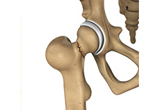
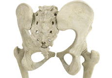
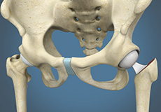
The hip joint is the junction where the hip joins the leg to the trunk of the body. It is comprised of two bones: the thigh bone or femur and the pelvis which is made up of three bones called ilium, ischium, and pubis. The ball of the hip joint is made by the femoral head while the socket is formed by the acetabulum. The Acetabulum is a deep, circular socket formed on the outer edge of the pelvis by the union of three bones: ilium, ischium and pubis. The lower part of the ilium is attached by the pubis while the ischium is considerably behind the pubis. The stability of the hip is provided by the joint capsule or acetabulum and the muscles and ligaments which surround and support the hip joint.
The head of the femur rotates and glides within the acetabulum. A fibrocartilaginous lining called the labrum is attached to the acetabulum and further increases the depth of the socket.
The femur or thigh bone is one of the longest bones in the human body. The upper part of the thigh bone consists of the femoral head, femoral neck, and greater and lesser trochanters. The head of the femur joins the pelvis (acetabulum) to form the hip joint. Next to the femoral neck, there are two protrusions known as greater and lesser trochanters which serve as sites of muscle attachment.
Articular cartilage is the thin, tough, flexible, and slippery surface lubricated by synovial fluid that covers the weight-bearing bones of the body. It enables smooth movements of the bones and reduces friction.
Ligaments
Ligaments are fibrous structures that connect bones to other bones. The hip joint is encircled with ligaments to provide stability to the hip by forming a dense and fibrous structure around the joint capsule. The ligaments adjoining the hip joint include:
- Iliofemoral ligament – This is a Y-shaped ligament that connects the pelvis to the femoral head at the front of the joint. It helps in limiting over-extension of the hip.
- Pubofemoral ligament – This is a triangular shaped ligament that extends between the upper portion of the pubis and the iliofemoral ligament. It attaches the pubis to the femoral head.
- Ischiofemoral ligament – This is a group of strong fibers that arise from the ischium behind the acetabulum and merge with the fibers of the joint capsule.
- Ligamentum teres – This is a small ligament that extends from the tip of the femoral head to the acetabulum. Although it has no role in hip movement, it does have a small artery within that supplies blood to a part of the femoral head.
- Acetabular labrum – The labrum is a fibrous cartilage ring which lines the acetabular socket. It deepens the cavity increasing the stability and strength of the hip joint.
Muscles and Tendons
A long tendon called the iliotibial band runs along the femur from the hip to the knee and serves as an attachment site for several hip muscles including the following:
- Gluteals – These are the muscles that form the buttocks. There are three muscles (gluteus minimus, gluteus maximus, and gluteus medius) that attach to the back of the pelvis and insert into the greater trochanter of the femur.
- Adductors – These muscles are in the thigh which help in adduction, the action of pulling the leg back towards the midline.
- Iliopsoas: This muscle is in front of the hip joint and provides flexion. It is a deep muscle that originates from the lower back and pelvis, and extends up to the inside surface of the upper part of the femur.
- Rectus femoris – This is the largest band of muscles located in front of the thigh. They also are hip flexors.
- Hamstring muscles – These begin at the bottom of the pelvis and run down the back of the thigh. Because they cross the back of the hip joint, they help in extension of the hip by pulling it backwards.
Nerves and arteries
Nerves of the hip transfer signals from the brain to the muscles to aid in hip movement. They also carry the sensory signals such as touch, pain, and temperature back to the brain.
The main nerves in the hip region include the femoral nerve in the front of the femur and the sciatic nerve at the back. The hip is also supplied by a smaller nerve known as the obturator nerve.
In addition to these nerves, there are blood vessels that supply blood to the lower limbs. The femoral artery, one of the largest arteries in the body, arises deep in the pelvis and can be felt in front of the upper thigh.
Hip movements
All the anatomical parts of the hip work together to enable various movements. Hip movements include flexion, extension, abduction, adduction, circumduction, and hip rotation.
Osteoarthritis of the Hip
Osteoarthritis, also called degenerative joint disease, is the most common form of arthritis. It occurs most often in older people. This disease affects the tissue covering the ends of bones in a joint (cartilage). In a person with osteoarthritis, the cartilage becomes damaged and worn out causing pain, swelling, stiffness and restricted movement in the affected joint.
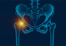
Hip Pain
Hip pain, one of the common symptoms patients complain of, may not always be felt precisely over the hip joint. Pain may be felt in and around the hip joint and the cause for pain is multifactorial. The exact position of your hip pain suggests the probable cause or underlying condition causing pain.
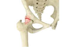
Femoroacetabular Impingement
Femoroacetabular impingement (FAI) is a condition where there is too much friction in the hip joint from bony irregularities causing pain and decreased range of hip motion. The femoral head and acetabulum rub against each other creating damage and pain to the hip joint.
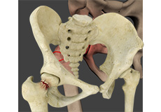
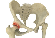
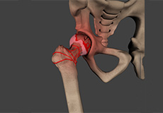
Hip Fracture
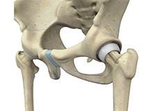
The hip joint is a “ball and socket” joint. The “ball” is the head of the femur, or thigh bone, and the “socket” is the cup shaped acetabulum. The joint surface is covered by a smooth articular surface that allows pain free movement in the joint.
Avascular Necrosis
Avascular necrosis, also called osteonecrosis is a condition in which bone death occurs because of inadequate blood supply to it. Lack of blood flow may occur when there is a fracture in the bone or a joint dislocation that may damage nearby blood vessels. Chronic use of high doses of steroid medications and heavy alcohol consumption are the two main risk factors of avascular necrosis.
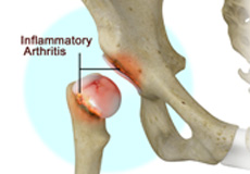
Inflammatory Arthritis of the Hip
Inflammation of the joints is referred to as arthritis. The inflammation arises when the smooth covering (cartilage) at the end surfaces of the bones wears away. In some cases, the inflammation is caused when the lining of the joint becomes inflamed as part of an underlying systemic disease. These conditions are referred to as inflammatory arthritis.
Non-Surgical Treatments
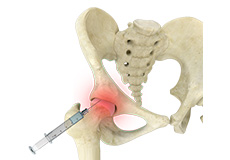
Hip Injections
Hip joint injections involve injecting medicine directly into the hip joint to diagnose the source of pain or treat pain due to conditions such as arthritis, injury or mechanical stress of the hip joint. Hip pain may be experienced in the hip, buttock, leg or low back.

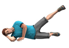
Physical Therapy
Physical therapy is an exercise program that helps you improve movement, relieve pain, encourage blood flow for faster healing, and restore your physical function and fitness level.
Surgical Treatment
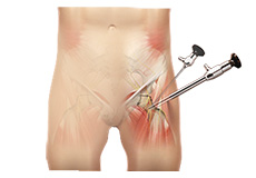
Hip Arthroscopy
Arthroscopy, also referred to as keyhole or minimally invasive surgery, is a procedure in which an arthroscope is inserted into a joint to check for any damage and repair it simultaneously.
Hip Resurfacing
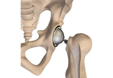
The hip joint is also known as a ball and socket joint, where the ball (femoral head) of the thigh bone fits into the socket (acetabulum) of the pelvis bone.
Damage to the hip bones can be treated by hip resurfacing, which is a surgical procedure in which the damaged parts of the femoral head are trimmed, and the socket is removed and replaced with metal caps.
Total Hip Replacement
Arthritis is inflammation of the joints resulting in pain, swelling, stiffness and limited movement. Hip arthritis is a common cause of chronic hip pain and disability.
Revision Hip Replacement
Revision hip replacement is a complex surgical procedure in which all or part of a previously implanted hip-joint is replaced with a new artificial hip-joint. Total hip replacement surgery is an option to relieve severe arthritis pain that limits your daily activities. During total hip replacement, the damaged cartilage and bone is removed from the hip joint and replaced with artificial components.
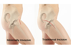
Minimally Invasive Total Hip Replacement
The hip joint is one of the body's largest weight-bearing joints and is the point where the thighbone (femur) and the pelvis (acetabulum) join. It is a ball and socket joint in which the head of the femur is the ball and the pelvic acetabulum forms the socket. The joint surface is covered by a smooth articular cartilage that cushions and enables smooth movements of the joint.
Outpatient Hip Replacement
Hip replacement surgery is the most common orthopedic surgery performed. It involves the replacement of the damaged hip bone (ball shaped upper end of the femur) with a metal ball attached to a metal stem that is fixed into the femur and attached to the pelvic region.
SuperPATH Hip Replacement
Supercapsular Percutaneously-Assisted Total Hip surgery or SuperPATH® surgery is a novel method of hip replacement where your surgeon can perform total hip replacement through 2-3-inch incision into the tip of the hip and without dislocating the hip or damaging the surrounding soft-tissue (muscles and tendons). This technique is also referred to as the Northern Approach or Northern Exposure.
Posterior Hip Replacement
Arthritis is a condition in which the articular cartilage that covers the joint surface is damaged or worn out causing pain and inflammation. Some of the causes of arthritis include:
Hip Hemiarthroplasty
The hip joint is one of the body's largest weight-bearing joints and is the point where the thighbone (femur) and the pelvis (acetabulum) unite. It is a ball and socket joint in which the head of the femur is the ball and the pelvic acetabulum forms the socket. The joint surface is covered by a smooth articular cartilage that cushions and enables smooth movements of the joint.
Pre-op and Post-op Hip Guidelines
Planning for your hip surgery prepares you for the operation and helps to ensure a smooth surgery and easier recovery. Here are certain pre-operative and post-operative guidelines which will help you prepare for hip surgery.
Hip Fracture Prevention
Hip fractures refer to any kind of breakage or damage in the thighbone (femur). People over the age of 65, especially women, are highly vulnerable to hip fractures. You will require assistance after hip fractures from family members as well as health professionals and may also be required to be admitted to the hospital for further assistance.